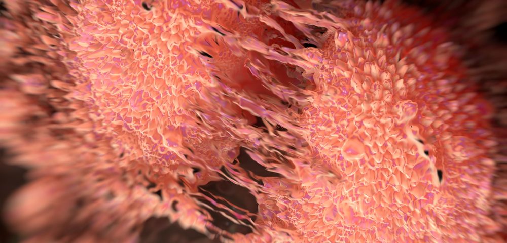
How prostate cancer turns metastatic and begins to spread — the first stage in what can become a deadly disease — takes place through a process not fully understand in this or any cancer. But researchers in the U.K. developed a cell model for prostate cancer that may help to unravel what is now a bit of a mystery.
The study, “A novel spontaneous model of epithelial-mesenchymal transition (EMT) using a primary prostate cancer derived cell line demonstrating distinct stem-like characteristics,” was published in the journal Scientific Reports.
The process that allows cancer cells to move from a stationary state, in which they are tightly attached to neighboring cells, into a migratory one — in which cells loosen connections, change shape, and start invading nearby tissues — is called the epithelial to mesenchymal transition (EMT).
Similar to many other cancers, EMT is believed to play a critical role in the spread of prostate cancer, but the lack of models has hindered research into the metastatic process.
Indeed, cell lines for prostate cancer research are still being derived from prostate cancer metastases, as it has been difficult to establish cell lines from a primary tumor. “To our knowledge, we are the first to derive and interrogate a spontaneous model of prostate cancer EMT,” the researchers said.
“Cancer cells acquire the capacity to move from the primary tumour to other sites by activating biological processes which allow them to survive the journey and establish themselves in their new ‘home,'” Dr. David Boocock, a scientist in Nottingham Trent University’s John van Geest Cancer Research Centre, said in a press release. “It is clear that understanding these processes is crucial if we are to reduce the number of prostate cancer-related deaths.”
Researchers at the van Geest center hypothesized that cell lines derived from a primary tumor site might contain information important in the onset of the metastatic process. They used less common primary tumor-derived cell lines to develop unique subpopulations of cells derived from those lines to serve as a model.
Experiments showed that two of these four subpopulations were able to spontaneously undergo EMT, while the other two could not. One of these two EMT-active subpopulations, called P4B6, also exhibited aggressive stem cell-like properties.
This suggests that not all tumor cells with the potential to migrate can initiate a metastasis in other parts of the body, while others naturally took on metastatic and mobile features. The researchers highlight that this model, which helps to explain the metastatic process before it begins, could support other studies into the mechanistic process of EMT and metastasis in prostate cancer.
“The model developed in the present work provides a valuable resource for the investigation of EMT in human prostate cancer. Unlike artificially-induced models of EMT, the EMT-derived cells in this study express endogenous levels of EMT-associated proteins. As such, these clones can be utilised to derive an authentic EMT-signature, which could be extended to clinical applications,” the researchers concluded.
Added Graham Pockley, director of the van Geest Cancer Research Centre: “This work provides a novel and important platform for future studies that will help us to predict prostate cancer metastasis and better understand cancer progression. As such, it could also be crucial in providing valuable insight into potential new therapies and approaches for the treatment and management of prostate cancer.”

