Metal tags enabled identification of cancer cells in a patient with metastatic prostate cancer, according to a study that suggests the method could provide new understanding of how cancer spreads and help define personalized treatments.
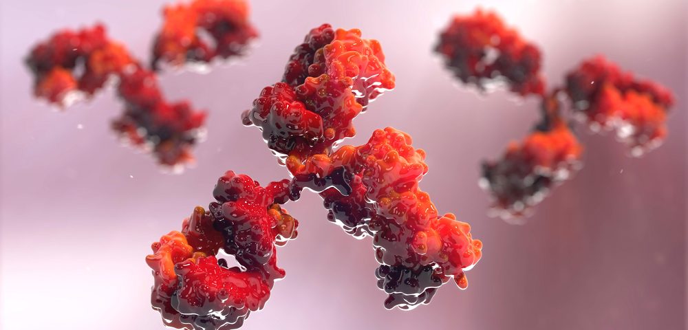
Using blood and bone marrow samples from the patient, researchers from the Bridge Institute at the University of Southern California (USC) applied metal-tagged antibodies targeting potential protein biomarkers, a technique called mass cytometry. This approach led to digital facsimiles of tumor cells that travel through the body.
“That is exactly what is happening when the TSA [Transportation Security Administration] swipes your hands,” Peter Kuhn, PhD, the study’s senior author and a professor of biological sciences at USC, said in a press release. “They are looking for metals, which are really easy to identify.”
The research, “Multiplex protein detection on circulating tumor cells from liquid biopsies using imaging mass cytometry,” was published in the journal Convergent Science Physical Oncology.
Investigators aimed to obtain an improved blueprint for the spread of the tumor, which is an advanced stage in cancer with challenging treatment options. Circulating tumor cells break away from the original tumor and may infiltrate into organs or bones, where they metastasize.
“We are trying to understand how cancer actually moves from the initial location to other places in the body and can settle there,” Kuhn said.
Using an imaging system to monitor cancer cells, scientists could see protein biomarkers at a molecular level not achieved before. Investigators had been relying on fluorescence microscopy, which images fluorescent-labeled antibodies, but the limited number of colors it provides restrains its use.
Altogether, the technique enabled the simultaneous tracking of 35 different metal labels, which means 35 distinct views of the cancer cell’s biology, Kuhn said.
“Oftentimes, we sequence the cancer’s genetic code, and that’s great because the only way to build something like a building or a machine is with a blueprint. But not every blueprint ends up being built to specification or even perform as expected,” Kuhn added.
“For a closer perspective and for purposes of improving the precision of medical treatment, you have to move in, from genome to proteome to cell,” he commented.
Zooming in on circulating and disseminated cancer cells would then provide deeper knowledge of the cells’ behavior. Importantly, the metal-tracing strategy also enables monitoring the course of treatment and evaluate changes in the patient’s immune system.
A 2013 study from the University of Zurich, Switzerland, first laid the foundations for using metals to characterize cancer. The USC team now expands on this work by using a liquid biopsy approach, which uses a simple blood sample.
“We simply add the antibody cocktail, wait a while for binding and then wash off the excess and see what sticks — like tie dye,” Kuhn said.
“While the technology is currently applied as a discovery tool for markers or new combinations of markers, it has the potential for clinical validity either as a broader pathology platform, or during the development phase of a specific test,” researchers wrote.
Due to its positive results, the method is now an official product of Fluidigm and can be used by researchers worldwide.
“This is really just the beginning,” Kuhn added. “You’ll see hundreds of studies now using this technique.”

