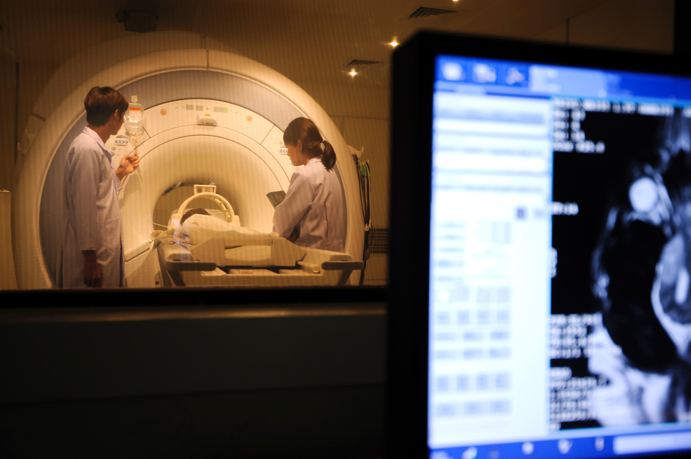
 A research team has developed an improved MRI technique that can provide more effective and precise treatments to address prostate cancer. This imaging technique allows doctors and imaging professionals to obtain clearer images of the precise location of the tumor and its severity. Thanks to a sharper image, more accurate biopsies can be executed, better treatment planning is enabled and surgeons are able to better pinpoint a tumor without affecting surrounding healthy tissue.
The research team included scientists from the University of California, San Diego and the University of California, Los Angeles, and their results have been published in the Prostate Cancer and Prostatic Disease journal.
Prostate cancer is the number-one cancer affecting men, aside from skin cancer. This research study addresses a crucial issue in men’s health. The American Cancer Society estimates over 220,800 new cases of prostate cancer and nearly 27,540 deaths resulting from the disease in 2015 alone.
This new approach allows the restriction of spectrum imaging MRI (RSI-MRI) and improves on the currently used diffusion MRI techniques by correcting for magnetic field distortion to give a more exact image of tumor position.
“Current imaging of prostate cancer is done with contrast-enhanced MRI. Unfortunately, some tumors fail to show a marked difference from surrounding healthy tissue due to lack of uptake of the contrast agent.” The diffusion MRI technique improves contrast MRI, however, magnetic field artifacts often distort tumor location. “RSI-MRI corrects for this distortion, improving the accuracy of MRI to localize tumors,” explained Nate White from UCSD.
In this study, 9 prostate cancer patients were assessed with both RSI-MRI and standard MRI. Researchers observed significant results for RSI-MRI in identifying extraprostatic extension (EPE).
More clinical testing is needed to understand the value of this technique and the best way to use it; it is important to assess its capacity of identifying adequate EPE treatment plans for these patients suffering with aggressive cancers.
“And, in cases where surgery is warranted, the accurate RSI-MRI image will also guide a more discriminating surgery to completely remove the tumor while sparing surrounding healthy tissues; this is an important consideration for allowing patients to maintain sexual function and urinary control following surgery,” concluded David S. Karow, UCSD.

