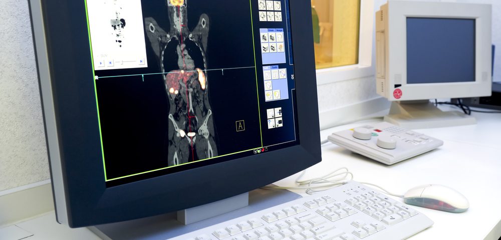
SmartTarget, an image fusion software that combines magnetic resonance imaging (MRI) and ultrasounds, should be used along with a visual review of MRI scans to conduct biopsies in patients with suspected prostate cancer, a study shows.
The study, “The SmartTarget Biopsy Trial: A Prospective, Within-person Randomised, Blinded Trial Comparing the Accuracy of Visual-registration and Magnetic Resonance Imaging/Ultrasound Image-fusion Targeted Biopsies for Prostate Cancer Risk Stratification,” was published in European Urology.
Prostate cancer diagnosis depends on accurate identification of the tumor during an invasive biopsy. Until recently, doctors performed a biopsy using an ultrasound, without knowing the location of the tumor. But the reliability of the approach was low and many tumors were not detected.
In recent years, the addition of MRI imaging — which indicates the most likely location of the tumor before performing the biopsy — has shown good results, increasing the percentage of identified tumors and making the procedure less invasive.
“Prostate cancer detection has been improving at a very fast rate in recent years, and this technology pushes the science forward even further, enabling clinicians to pick up prostate cancer quickly so that patients can access the right treatment early enough,” professor Hashim Ahmed, co-senior author of the study, said in a press release.
MRI-guided biopsies require an experienced surgeon who can translate MRI findings into what he is seeing on the ultrasound during a biopsy, a strategy called traditional visual registration.
To facilitate the procedure, a team of engineers and medical researchers at University College London developed SmartTarget, software that helps locate tumors by fusing the MRI imaging and the live ultrasound, a strategy called image-fusion.
“We developed the SmartTarget system to equip surgeons with vital information about the size, shape, and location of prostate tumors during a biopsy that is otherwise invisible on ultrasound images,” said co-senior author Dean Barratt, who invented and led the development of the SmartTarget system.
The SmartTarget Biopsy trial (NCT02341677) was designed to compare the efficacy of SmartTarget-guided biopsies to the visual registration biopsies in detecting prostate cancer.
The study enrolled 129 men with suspected prostate cancer determined by MRI. Each patient underwent both kinds of biopsies and the results were compared.
When combined, the two strategies detected 93 clinically significant prostate cancers — each approach identified 80 cases and missed 13 cancers detected by the other. Because the results were similar, researchers recommended combining strategies.
However, the surgeons in this study were highly skilled, and researchers believe that SmartTarget is likely to outperform visual registration biopsies conducted by less experienced doctors.
“We hope that the imagery displayed by SmartTarget will help to bring high-accuracy prostate cancer diagnosis to a much wider range of patients and hospitals,” Barratt said.
The software is commercialized by SmartTarget; several hospitals in the U.K. and U.S. are already using it.
“There has been much discussion and speculation in the media recently on the degree to which computers and artificial intelligence will be integrated into clinical care. Studies such as this one are extremely important as they provide valuable evidence on the performance of a new technology in the clinical setting,” said professor Mark Emberton, co-senior author.
“With this study, we now have hard data showing that SmartTarget is as good as a group of experts in targeting tumors in the prostate, and have a glimpse of how clinicians and computers will be working together in the future for the good of the patient.”

