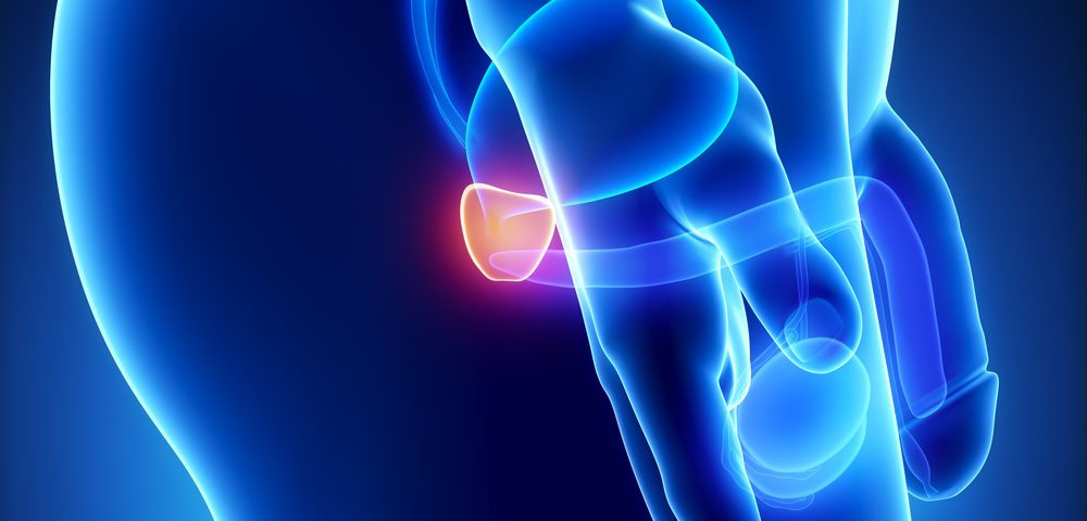
A research team at the Thayer School of Engineering at Dartmouth received a $2.5 million grant to develop and test a device meant to help surgeons detect and remove prostate cancer cells that would otherwise be missed during surgery.
The five-year grant, awarded by the National Cancer Institute, part of the National Institutes of Health, will focus on differences in an electric current when it flows through cancer cells, according to a University press release.
The complete removal of a tumor during surgery helps to avoid additional surgeries and treatments, and cancer relapse. Most importantly, it helps increase patients’ overall survival.
Distinguishing between cancer and healthy tissue during surgery currently depends mainly on visual inspection and palpation by the surgeon — a method with undetermined accuracy. Surgeons routinely remove an extra centimeter of tissue around a tumor to reduce the chances of leaving any cancer cells behind.
Still, according to a 2014 review study, 6.5% to 32% of men undergoing radical prostatectomy each year come out of surgery with positive surgical margins. That means that cancer cells were still detected at the edges of the removed tissue, according to pathology reports.
The presence of positive surgical margins increases the likelihood that some cancer may have been left behind, and many patients often choose to undergo radiation therapy, hormone therapy, or chemotherapy to eradicate the leftover disease. Such additional therapies can increase a patient’s physical and financial burden.
Therefore, there is an unmet need to improve the specific detection of tumor margins during surgical procedures.
The Dartmouth team, led by Ryan Halter, PhD, a professor of engineering and surgery, will focus on the development and testing of a small device — called a microendoscopic electrical impedance sensing probe — that can distinguish cancer cells from healthy ones by sensing differences in electric current flow.
In previous studies, Halter and his colleagues showed that the electrical properties of prostate cancer tissue are distinct from those of non-cancer prostate tissue. His team found that these differences can be used to effectively discriminate between the two tissue types.
Halter previously developed an early prototype of the device — for which he also won a grant award — at his company, RyTek Technical Service, which focuses on translating the technologies developed in his lab to the market. With this new grant, the team will be able to study the device in the first large study in humans.
“We’re really trying to augment and improve a patient’s quality of life following surgery, and intraoperatively detecting positive surgical margins will help to make that a reality,” Halter said.
“One of the things this approach might allow is for surgeons to be more aggressive in nerve-sparing procedures because they can check that margin after they’ve done the procedure to see if there’s cancer left behind,” he added.
During the grant’s first year, the team — which includes Dartmouth engineering and medical colleagues and students — will optimize the device, which will be miniaturized and integrated within a surgical probe to allow for a better signal and clearer data.
Starting in the second year, the device will be tested in 200 men with prostate cancer undergoing radical prostatectomy. In the operating room, members of the Dartmouth team will collect data in real time to identify potential positive surgical margins while a surgeon manipulates the probe with the device.
Lawrence Dagrosa, part of the team and a professor of surgery and specialist in urological oncology at Dartmouth-Hitchcock Medical Center, said that “currently, there is no way to test for the presence of surgical margins in the operating room. The ability to identify, and subsequently address, positive surgical margins in real-time will be a game-changing advancement in the surgical treatment of prostate cancer.”
Halter believes the device will be especially helpful to patients with larger positive surgical margins, or tumors largely going beyond the removed tissue, who are more likely to have poorer quality of life.
He noted, however, that the tool may not have the same resolution level as the current method of assessing the presence of positive surgical margins, which involves microscopic analysis of the removed tissue.
Should the pivotal human studies be successful, Halter expects to conduct larger clinical trials and explore the device’s potential in surgeries for other cancer types, such as those affecting the breast, kidneys, and brain.

